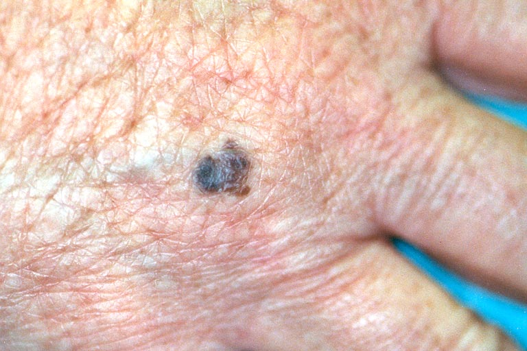
A pigmented lesion measuring 5 mm in diameter is present on the dorsum of the right hand. There is modest asymmetry, and a subtle notch in the border at the medial side of the lesion. No variegation of color is present.
![]()
A 52-year-old woman presented with a pigmented papule on the dorsum of the right hand, measuring 5 mm in diameter. The lesion had a homogenous brown-black color, "stuck-on" appearance, with only slight border irregularity, and asymmetry (FIG. 1).
|
Figure 1
A pigmented lesion measuring 5 mm in diameter is present on the dorsum of the right hand. There is modest asymmetry, and a subtle notch in the border at the medial side of the lesion. No variegation of color is present. |
|
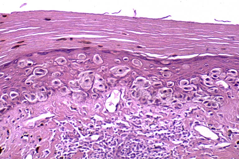
The primary diagnosis in the clinical differential diagnosis was seborrheic keratosis. The patient's report of a recent change in size and darkening pigmentation also prompted punch excision. Histopathologic investigation showed irregular proliferation of atypical melanocytes showing variation in nuclear shape, size, and chromatin pattern at the dermal-epidermal junction, with pagetoid infiltration of the epidermis and dermal invasive growth to a depth of 0.20 mm (Breslow) Clark's Level II (FIGS. 2a,2b,2c).
|
Figure 2a
A scanning magnification view showing pagetoid melanocytes, intradermal nests of melanocytes, chronic inflammation and scattered melanophages. |

|
|
Figure 2b
A close-up view showing the extensive pagetoid in situ component of this melanoma. |
|
|
Figure 2c
A close-up view of another area of this melanoma highlighting an invasive nest of melanoma in addition to the in situ component. Although not definitive, vascular invasion is suggested. |

|
The diagnosis of superficial spreading malignant melanoma was made.
© 1999 Dermatology Online Journal