Case 5
A 39 year-old man presented with a three month history of a tender, enlarging mole on the right medial thigh. Physical examination
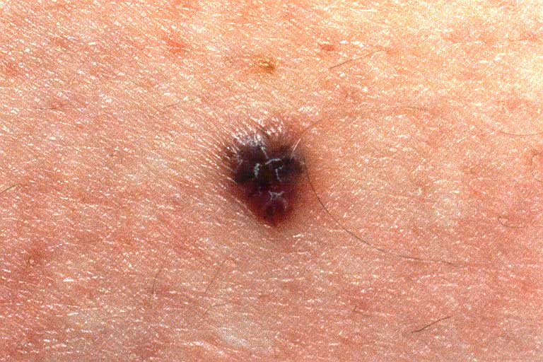
revealed a symmetric, dark-brown nodule, 3 mm in diameter with a regular border and a slight rim of erythema (Figure 9).
|
Figure 9
A pigmented, 3 mm nodule of the right medial thigh, with a slight rim of erythema.
Photograph used with permission of Postgraduate Medicine, from Brodell RT, Helms SE, The changing mole: Additional warning
signs of malignant melanoma, Postgrad Med, 104(4):145-148, October 1998. C 1988 The McGraw-Hill Companies.
|

|
Clinically it was felt to be an inflamed benign melanocytic nevus. Because it was enlarging, a punch biopsy was performed
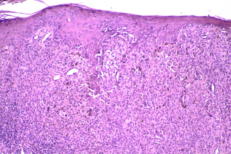
which demonstrated irregular masses of atypical melanocytes arising from the dermal-epidermal junction and invading the dermis
to a depth of 1.39 mm, Clark's Level IV (Figures 10a, 10b).
|
Figure 10a
A scanning magnification view showing a primarily invasive melanoma.
|
|
|
Figure 10b
A close-up view showing both the in situ as well as the invasive components of the melanoma.
|

|
The lesion was re-excised, but two years later the patient developed right inguinal lymphadenopathy, which was found to contain
malignant melanoma. He died of widespread multi-system malignant melanoma within 18 months.
© 1999 Dermatology Online Journal