

Tropical Endomyocardial Fibrosis: Followed by a case of Endomyocardial Fibrosis by Christine McBeth, DO, MSPH
Abstract
A 45-year-old male arrives to our mobile clinic after walking from his village 20km away with three days of a worsening fever, myalgias, and fatigue. He has not eaten in 48 hours. The walk from his village proved extremely difficult for him and he noted also feeling mildly dyspneic...
Main Content
Endomyocardial Fibrosis
Christine McBeth, DO, MSPH
Department of Emergency Medicine, University of California Davis
Clinical Presentation
History
42-year old-female from Uganda presented with shortness of breath for several months that acutely became worse over the past two days. She has a history of unknown cardiac disease and had undergone a pericardiocentesis two months prior that had improved some of her symptoms. She has had progressive weight gain, edema, cough and shortness of breath.
Physical exam
| Blood pressure | Pulse | Respiratory Rate | Pulse Oximetry | Temperature |
|---|---|---|---|---|
| 65/30 | 145 | 48 | Unknown | 98.9 |
General:
Ill-appearing, tachypneic, obtunded
Cardiovascular:
Tachycardic, irregularly irregular, no murmurs, +JVD
Respiratory:
Sitting up in tripod position, tachypneic, diffuse crackles
Imaging and Laboratory Information
Chest X-ray: normal heart size, diffuse pulmonary edema, no pleural effusions or infiltrates
Clinical Differential Diagnosis
- Congestive heart failure
- Pneumonia
- Rheumatic heart disease
- Tuberculosis
- Myocardial infarction
- Tamponade
- Pulmonary embolism
- Endomyocardial fibrosis
Ultrasound Imaging Findings
Key:
RA: right atrium
LA: left atrium
RV: right ventricle
LV: left ventricle
PCE: pericardial effusion
IVC: inferior vena cava
**: intra-atrial thrombus
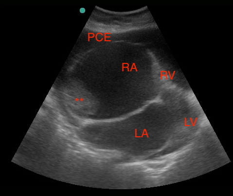 Subxiphoid view: small-moderate pericardial effusion, markedly enlarged right atrium and left atrium, clot present right atrium, no right atrial collapse, barely visible right and left ventricles (constricted).
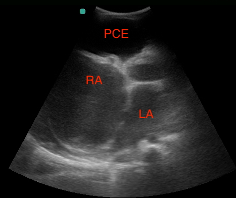 Parasternal short: moderate pericardial effusion, markedly enlarged right atrium, unable to visualize right ventricle.
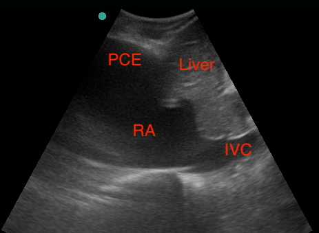 IVC: markedly enlarged right atrium with plethoric IVC.
Differential Diagnosis Based on Imaging
- Endomyocardial fibrosis
- Effusion with tamponade
- Tuberculous constrictive pericarditis
- Dilated cardiomyopathy
- Ventricular free wall rupture
Clinical Course and/or Management
The patient was placed on oxygen and given 80mg of IV furosemide. It was decided that given ultrasound findings, the patient was not in tamponade and pericardiocentesis was not performed. The patient was then started on "dirty epi drip" by placing 1mg epinephrine into a 1,000 mL normal saline bag wide open through 18 gauge peripheral IV and given broad spectrum antibiotics. The patient expired within 24 hours.
Diagnosis
Tropical endomyocardial fibrosis
Discussion
Tropical endomyocardial fibrosis (EMF) was first described in the 1940's and remains a complex and enigmatic cause of restrictive cardiomyopathy. The majority of cases are described clustered around the equator in Africa, with cases also described in Asia and South America. This disease is increased in those affected by poverty and has bimodal distribution at age 10 and 30 with very poor long-term outcomes, with approximately 75% mortality at two years. The cause of EMF is unclear, but a multi-factorial causation has been suggested that includes contributions from poverty and protein malnutrition, parasitic infection and eosinophilia, genetics and autoimmunity. The acute phase is associated with inflammation and high levels of eosinophilia, but is hard to diagnose and detect. The chronic phase has symptoms such as peripheral edema, ascites and signs of malnutrition and is characterized by endocardial fibrosis in the ventricles as well as the atrioventricular valves, which can lead to mitral and tricuspid regurgitation. Valvular abnormalities can cause dilation of the atria, which can lead to atrial fibrillation and thromboses. Echocardiography is the standard diagnostic modality with findings of thickened endocardium, severely dilated atria, atrioventricular valve dysfunction, retracted ventricles and often apical thrombi. This can be differentiated from tuberculosis associated constrictive pericarditis by having a normal pericardial thickness. Treatment options include steroids for anti-inflammatory benefits, although these have not been shown to improve survival, symptomatic heart failure treatment, anticoagulants and surgical management such as endocardectomy and valvular repair if available.
References
- Grimaldi A, Mocumbi A, Freers J, et al. Tropical Endomyocardial Fibrosis: Natural History, Challenges, and Perspectives. J Circulation 2016; 133: 2503-2515.
- Beaton A and Mocumbi A. Diagnosis and Management of Endomyocardial Fibrosis. Cardiology Clinics 2017; 35(1): 87-98.